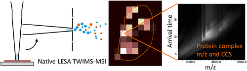
We have previously demonstrated native liquid extraction surface analysis (LESA) mass spectrometry imaging of small intact proteins in thin tissue sections. We also showed calculation of collision cross sections for specific proteins extracted from discrete locations in tissue by LESA traveling wave ion mobility spectrometry (TWIMS). Here, we demonstrate an integrated native LESA TWIMS mass spectrometry imaging (MSI) workflow, in which ion mobility separation is central to the imaging experiment and which provides spatial, conformational, and mass information on endogenous proteins in a single experiment. The approach was applied to MSI of a thin tissue section of mouse kidney. The results show that the benefits of integration of TWIMS include improved specificity of the ion images and the capacity to calculate collision cross sections for any protein or protein complex detected in any pixel (without a priori knowledge of the presence of the protein).
Authors: Cecilie Rosting, Jinglei Yu, and Helen J. Cooper*
Abstract
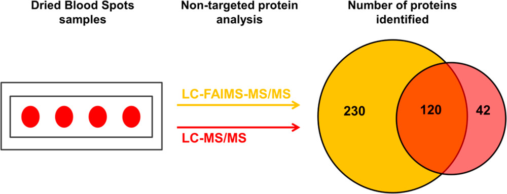
Despite the great potential of dried blood spots (DBS) as a source of endogenous proteins for biomarker discovery, the literature relating to nontargeted bottom-up proteomics of DBS is sparse, primarily due to the inherent complexity and very high dynamic range associated with these samples. Here, we present proof-of-concept results in which we have coupled high field asymmetric waveform ion mobility spectrometry (FAIMS) with liquid chromatography–tandem mass spectrometry (LC–MS/MS) for nontargeted bottom-up proteomics of DBS with the aim of addressing these challenges.
We, and others, have previously demonstrated the benefits of FAIMS more generally in proteomics including improved signal-to-noise and extended proteome coverage, and the aim of the current work was to extend those benefits specifically to DBS. The DBS samples were either extracted by the more traditional manual “punch and elute” approach or by an automated liquid surface extraction (LESA) approach prior to trypsin digestion. The resulting samples were analyzed by LC–MS/MS and LC–FAIMS–MS/MS analysis. The results show that the total number of proteins identified increased by ∼50% for the punch and elute samples and ∼45% for the LESA samples in the LC–FAIMS–MS/MS analysis. For both the punch and elute samples and the LESA samples, ∼30% of the total proteins identified were observed in both the LC–MS/MS and the LC–FAIMS–MS/MS data sets, illustrating the complementarity of the approaches.
Overall, this work demonstrates the benefits of inclusion of FAIMS for nontargeted proteomics of DBS.
Liquid extraction surface analysis (LESA) is an ambient surface sampling technique that allows the analysis of intact proteins directly from tissue samples via mass spectrometry. Integration of ion mobility separation to LESA mass spectrometry workflows has shown significant improvements in the signal-to-noise ratios of the resulting protein mass spectra and hence the number of proteins detected. Here, we report the use of a quadrupole–cyclic ion mobility–time-of-flight mass spectrometer (Q-cIM-ToF) for the analysis of proteins from mouse brain and rat kidney tissues sampled via LESA. Among other features, the instrument allows multiple pass cyclic ion mobility separation, with concomitant increase in resolving power. Single-pass experiments enabled the detection of 30 proteins from mouse brain tissue, rising to 44 when quadrupole isolation was employed. In the absence of ion mobility separation, 21 proteins were detected in rat kidney tissue including the abundant α- and β-globin chains from hemoglobin. Single-pass cyclic ion mobility mass spectrometry enabled the detection of 60 additional proteins. Multipass experiments of a narrow m/z range (m/z 870–920) resulted in the detection of 24 proteins (one pass), 37 proteins (two passes) and 54 proteins (three passes), thus demonstrating the benefits of improved mobility resolving power.
Helen J. Cooper, Emma K. Sisley, Jakub Ujma, Martin Palmer, Kevin Giles, Francisco A. Fernandez-Lima
Daniel J. Ryan, David Nei, Boone M. Prentice, Kristie L. Rose, Richard M. Caprioli, Jeffrey M. Spraggins
Robotic liquid surface extractions can be used to interrogate discrete regions of tissue to provide protein identifications with high throughput, accuracy, and robustness. The direct coupling of tissue surface extractions and liquid chromatography, offers a new and effective approach to provide spatial proteomics data in an imaging experiment.
Tissue extractions were completed using the TriVersa NanoMate® (Advion, Inc., Ithaca, NY, USA) modified to include a glass capillary (LESA Plus) for improved spatial resolution and online integration with LC-based experiments. 36 Scanned images of thaw-mounted samples were uploaded to the ChipSoft Software (Advion, Inc.) to allow histological regions of interest to be selected for analysis.
Lieke Lamont, Mark Baumert, Nina Ogrinc Potočnik, Mark Allen, Rob Vreefken, Ron M. A. Heeren, and Tiffany Porta
Direct analysis by mass spectrometry (imaging) has become increasingly deployed in preclinical and clinical research due to its rapid and accurate readouts. However, when it comes to biomarker discovery or histopathological diagnostics, more sensitive and in-depth profi ling from localized areas is required. We developed a comprehensive, fully automated online platform for high-resolution liquid extraction surface analysis (HR-LESA) followed by micro−liquid chromatography (LC) separation and a data-independent acquisition strategy for untargeted and low abundant analyte identifi cation directly from tissue sections. Applied to tissue sections of rat pituitary, the platform demonstrated improved spatial resolution, allowing sample areas as small as 400 μ m to be studied, a major advantage over conventional LESA.
The LESA extraction was performed using the automated TriVersa NanoMate® The LESA extraction was controlled by a beta version of the LESA Plus software (Advion, UK)
Rian L. Griffiths, Emma K. Sisley, Andrea F. Lopez-Clavija, Anna L. Simmonds, Iain B. Styles, Helen J. Cooper
Here, researchers present native liquid extraction surface analysis (LESA) mass spectrometry imaging of proteins and protein complexes from mouse brain and liver tissue. Intact proteins were detected in characteristically low charge states, indicating that the proteins remain folded. In brain, abundant proteins such as ubiquitin and β thymosin 4 were detected homogeneously across the tissue whereas other proteins, such as neurogranin, were localised in specific anatomical regions.
In liver, imaging of a protein complex (tetrameric hemoglobin) is demonstrated, as well as fatty acid binding protein. Interestingly, the use of native-like solvents enables extraction of proteins which have not previously been observed in LESA experiments employing denaturing solvents, i.e., native LESA can be applied to extend the range of proteins observed. In addition native LESA ion mobility spectrometry is presented and shows that the collision cross sections of proteins extracted from tissue may be determined by travelling wave ion mobility spectrometry. The collision cross section of the 5+ ion of ubiquitin was calculated as 1047 Å2, in good agreement with measurements of ubiquitin protein standard solutions. Collision cross sections for the 4+ ions of β-thymosin 4, β-thymosin 10 and two unidentified proteins were also calculated, together with that of a 10+ ion of an unidentified protein of molecular weight 15660 Da.
For mass spectrometry and mass spectrometry imaging, the samples were introduced to the mass spectrometer via nanoESI using a TriVersa NanoMate® (Advion Biosciences, Ithaca, USA). The exact location to be sampled was selected using the LESA® Points software (Advion).
The TriVersa NanoMate® LESA® is the latest in chip-based electrospray ionization technology from Advion Interchim Scientific®. It combines the benefits of liquid chromatography, mass spectrometry, chip-based infusion, fraction collection and direct surface analysis into one integrated ion source platform. It allows scientists to obtain more information from complex samples than LC/MS alone.
Published by the International Journal of Mass Spectrometry
Native liquid extraction surface analysis (LESA) mass spectrometry enables the direct sampling of protein complexes from a solid surface. We have previously demonstrated native LESA mass spectrometry of holomyoglobin (~17 kDa) from glass slides and tetrameric haemoglobin (~64 kDa) from dried blood spots and thin tissue sections. Here, we further explore the capabilities of this
emerging technique by investigating a range of proteins which exist in various oligomeric states in vivo.
Victor A.Mikhailov, Rian L.Griffiths, Helen J.Cooper,
Liquid Extraction Surface Analysis for Native Mass Spectrometry: Protein
Complexes and Ligand Binding, International Journal of Mass Spectrometry
http://dx.doi.org/10.1016/j.ijms.2016.09.011