M. Montowska, M.R. Alexander, G.A. Tucker, D.A. Barrett
Anal Chem. 2014 Oct 21;86(20):10257-65. doi: 10.1021/ac502449w
In this Article, our previously developed ambient LESA-MS methodology is implemented to analyze five types of thermally treated meat species, namely, beef, pork, horse, chicken, and turkey meat, to select and identify heat-stable and species-specific peptide markers. In-solution tryptic digests of cooked meats were deposited onto a polymer surface, followed by LESA-MS analysis and evaluation using multivariate data analysis and tandem electrospray MS. The five types of cooked meat were clearly discriminated using principal component analysis and orthogonal partial least-squares discriminant analysis. 23 heat stable peptide markers unique to species and muscle protein were identified following data-dependent tandem LESA-MS analysis. Surface extraction and direct ambient MS analysis of mixtures of cooked meat species was performed for the first time and enabled detection of 10% (w/w) of pork, horse, and turkey meat and 5% (w/w) of chicken meat in beef, using the developed LESA-MS/MS analysis. The study shows, for the first time, that ambient LESA-MS methodology displays specificity sufficient to be implemented effectively for the analysis of processed and complex peptide digests. The proposed approach is much faster and simpler than other measurement tools for meat speciation; it has potential for application in other areas of meat science or food production.
The Liquid Extraction Surface Analysis (LESA) capability of the TriVersa NanoMate enables simple, direct nanoESI mass spectrometric analysis from a variety of surfaces.
The new LESAPLUS allows for automated LESA experiments plus additional nano-LC separation through the ChipSoftX operating software with Developers Kit. This enhancement is ideal for direct tissue analysis.
ChipSoftX is an entirely new operating software for the TriVersa NanoMate automated nanoelectrospray source. Besides improvement in program compatibility with Windows and integration of existing software features, it also provides access to the new Developers Kit – a platform for customized method development with direct access to robot controls allowing entirely novel analysis workflows such as LESAPLUS.
Native mass spectrometry seeks to probe noncovalent protein interactions in terms of protein quaternary structure, protein–protein and protein–ligand complexes. The ultimate goal is to link the understanding of protein interactions to the protein environment by visualizing the spatial distribution of noncovalent protein interactions within tissue. Previously, we have shown that noncovalently bound protein complexes can be directly probed via liquid extraction surface analysis from dried blood spot samples, where hemoglobin is highly abundant. Here, we show that the intact hemoglobin complex can be sampled directly from thin tissue sections of mouse liver and correlated to a visible vascular feature, paving the way for native mass spectrometry imaging.
R.L. Griffiths and H.J. Cooper Anal. Chem., 2016, 88 (1), pp 606–609
Rian Griffiths, Alex Dexter, Andrew Creese and Helen J Cooper Analyst, 2015, Accepted Manuscript DOI: 10.1039/C5AN00933B
Liquid extraction surface analysis (LESA) is a surface sampling technique that allows electrospray mass spectrometry analysis of a wide range of analytes directly from biological substrates. Here, we present LESA mass spectrometry coupled with high field asymmetric waveform ion mobility spectrometry (FAIMS) for the analysis of dried blood spots on filter paper. Incorporation of FAIMS in the workflow enables gas-phase separation of lipid and protein molecular classes, enabling analysis of both haemoglobin and a range of lipid (phosphatidylcholine or phosphatidylethanolamine, and sphingomyelin species) from a single extraction sample. The work has implications for multiplexed clinical assays of multiple analytes.
The search for therapeutic agents that bind specifically to precursor protein conformations and inhibit amyloid assembly is an important challenge. Identifying such inhibitors is difficult because many protein precursors of aggregation are partially folded or intrinsically disordered, which rules out structure-based design. Furthermore, inhibitors can act by a variety of mechanisms, including specific or nonspecific binding, as well as colloidal inhibition. Here we report a high-throughput method based on ion mobility spectrometry–mass spectrometry (IMS–MS) that is capable of rapidly detecting small molecules that bind to amyloid precursors, identifying the interacting protein species and defining the mode of inhibition. Using this method we have classified a variety of small molecules that are potential inhibitors of human islet amyloid polypeptide (hIAPP) aggregation or amyloid-beta 1-40 aggregation as specific, nonspecific, colloidal or non-interacting. We also demonstrate the ability of IMS–MS to screen for inhibitory small molecules in a 96-well plate format and use this to discover a new inhibitor of hIAPP amyloid assembly.
Randall EC, Bunch J, Cooper HJ; Anal Chem. 2014 Nov 4;86(21):10504-10. doi: 10.1021/ac503349d. Epub 2014 Oct 23
Top-down identification of proteins by liquid extraction surface analysis (LESA) mass spectrometry has previously been reported for tissue sections and dried blood spot samples. Here, we present a modified “contact” LESA method for top-down analysis of proteins directly from living bacterial colonies grown in Petri dishes,without any sample pretreatment. It was possible to identify a number of proteins by use of collision-induced dissociation tandem mass spectrometry followed by searches of the data against an E. coli protein database. The proteins identified suggest that the method may provide insight into the bacterial response to environmental conditions. Moreover, the results show that the “contact” LESA approach results in a smaller sampling area than typical LESA, which may have implications for spatial profiling.
Martin NJ, Griffiths RL, Edwards RL, Cooper HJ. J Am Soc Mass Spectrom. 2015 May 20. [Epub ahead of print]
Liquid extraction surface analysis (LESA) mass spectrometry is a promising tool for the analysis of intact proteins from biological substrates. Here, we demonstrate native LESA mass spectrometry of noncovalent protein complexes of myoglobin and hemoglobin from a range of surfaces. Holomyoglobin, in which apomyoglobin is noncovalently bound to the prosthetic heme group, was observed following LESA mass spectrometry of myoglobin dried onto glass and polyvinylidene fluoride surfaces. Tetrameric hemoglobin [(αβ)24H] was observed following LESA mass spectrometry of hemoglobin dried onto glass and polyvinylidene fluoride (PVDF) surfaces, and from dried blood spots (DBS) on filter paper. Heme-bound dimers and monomers were also observed. The ‘contact’ LESA approach was particularly suitable for the analysis of hemoglobin tetramers from DBS.
Our research focuses on in situ analysis of intact proteins from biological substrates. We combine ambient surface techniques and ion mobility spectrometry with high resolution mass spectrometry.
We are particularly interested in native ambient mass spectrometry, in which folded proteins, protein assemblies and protein complexes are sampled directly from thin tissue sections. Native ambient mass spectrometry, such as liquid extraction surface analysis (LESA), is integrated with mass spectrometry imaging to provide simultaneous spatial and structural information.
We also apply LESA for the analysis of intact but unfolded proteins from a range of substrates including living microbial colonies growing on agar and other solid substrates, dried blood spots and tissue sections. The combination of LESA, ion mobility spectrometry and mass spectrometry enables the detection of hundreds of proteins.
Why did you incorporate the TriVersa NanoMate® into your laboratory
Initially, we purchased the TriVersa NanoMate® for direct infusion and coupling to LC and we still use the equipment for that purpose. Advion Interchim Scientific’s chip technology has revolutionized nanospray as far as ease of use. The ESI Chip™ is robust and bypasses any problems with non-uniformity. It allows us to move simply to the next nozzle if there is an issue with spray. The spray sensing capability is very clever and necessary for our overnight runs. More recently, we have used the LESA capability of the TriVersa NanoMate® for our in situ analyses of proteins in tissue, dried blood spots and microbial colonies.
To whom would you recommend the TriVersa NanoMate® for their research?
I would recommend the TriVersa NanoMate® to anyone with a mass spectrometer who uses nanoelectrospray.
Do you have any publications or presentations using the TriVersa NanoMate®?
Publication Highlight
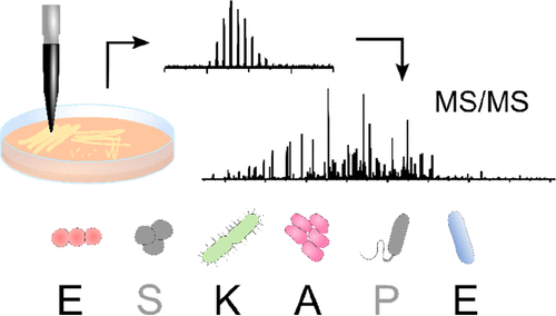
Liquid Extraction Surface Analysis Mass Spectrometry of ESKAPE Pathogens
Havlikova et al. J Am Soc Mass Spec. 2021
Top-down LESA MS/MS was used for protein identification in four ESKAPE pathogens as well as E. faecalis V583 and a clinical isolate of A. baumannii.
Other Publications:
Hale et al. Native mass spectrometry imaging and in situ top-down identification of intact proteins directly from tissue. J Am Soc Mass Spec. DOI: 10.1021/jasms.0c00226
Havlikova et al. Direct identification of bacterial and human proteins from infected wounds in living 3D skin models. Sci Rep. DOI: 10.1038/s41598-020-68233-6
Haque et al. Self-incompatibility triggers irreversible oxidative modification of proteins in incompatible pollen. Plant Physiology. DOI: 10.1104/pp.20.00066
Sisley et al. LESA cyclic ion mobility mass spectrometry of intact proteins from thin tissue sections. Anal. Chem. DOI: 10.1021/acs.analchem.9b05169
Hale et al. Native LESA TWIMS-MSI: Spatial, conformational, and mass analysis of proteins and protein complexes.J Am Soc Mass Spec. DOI: 10.1021/jasms.9b00122
Kocurek et al. Electroporation and mass spectrometry: A new paradigm for in situ analysis of intact proteins from living yeast colonies. Analytical Chemistry/ DOI: 10.1021/acs.analchem.9b04365
Griffiths et al. Comprehensive LESA mass spectrometry imaging of intact proteins by integration of cylindrical FAIMS. Analytical Chemistry. DOI: 10.1021/acs.analchem.9b05124
Havlikova et al. Quantitative imaging of proteins in tissue by stable isotope labeled mimetic liquid extraction surface analysis mass spectrometry. Analytical Chemistry. DOI: 10.1021/acs.analchem.9b04148
Griffiths et al. LESA MS imaging of heat-preserved and frozen tissue: Benefit of multistep static FAIMS. Analytical Chemistry. DOI: 10.1021/acs.analchem.8b02739
Rosting et al. High field asymmetric waveform ion mobility spectrometry in nontargeted bottom-up proteomics of dried blood spots. J Proteom Res. DOI: 10.1021/acs.jproteome.7b00746
Sarsby et al. Liquid extraction surface analysis mass spectrometry coupled with field asymmetric waveform ion mobility spectrometry for analysis of intact proteins from biological substrates. Analytical Chemistry. DOI: 10.1021/acs.analchem.5b01151
Griffiths et al. Liquid extraction surface analysis field asymmetric waveform ion mobility spectrometry mass spectrometry for the analysis of dried blood spots. Analyst. DOI: 10.1039/C5AN00933B
Sarsby et al. Top-down and bottom-up identification of proteins by liquid extraction surface analysis mass spectrometry of healthy and diseased human liver tissue. J Am Soc Mass Spec. DOI:10.1007/s13361-014-0967-z
Randall et al. Direct analysis of intact proteins from Escherichia coli colonies by liquid extraction surface analysis mass spectrometry. Analytical Chemistry. DOI: 10.1021/ac503349d
Edwards et al. Compound heterozygotes and beta‐thalassemia: Top‐down mass spectrometry for detection of hemoglobinopathies. PROTEOMICS. DOI: 10.1002/pmic.201300316
Martin et al. Dried blood spot proteomics: Surface extraction of endogenous proteins coupled with automated sample preparation and mass spectrometry. J Am Soc Mass Spec. DOI:10.1007/s13361-013-0658-1
Edwards et al. Hemoglobin variant analysis via direct surface sampling of dried blood spots coupled with high-resolution mass spectrometry. Analytical Chemistry. DOI: 10.1021/ac1030804
A: From drug-sized molecules to fragments, we analyze a little bit of everything. My primary research focuses around discovering the structure of proteins including enzyme structure and function in the development of antibiotics.
In addition, we have a collaboration with UCB, a global pharma focused on central nervous system and immunology disorders, involving library screening.
Q: Why did you incorporate the TriVersa NanoMate® into your laboratory?
A: We have a NanoMate 100 and a TriVersa NanoMate®. It is reasonably well-known that you need nanoESI for noncovalent interactions – ligand screening, but it was essential to my research to speed up and to automate the process. I had watched too many demonstrations that involved pulled capillaries, and I knew it would drive us crazy. A second instrument was absolutely vital to meet the needs of our collaboration with UCB.
Q: What benefits have you experienced with the TriVersa NanoMate®?
A: The main reason for purchasing the TriVersa NanoMate® is its automated nanoESI capability. The added benefit is its ease of use. We act as a service lab to the chemistry department, and often we have inexperienced users with a variety of different samples to be analyzed. The TriVersa NanoMate® makes it easy for users to do what needs to be done, very quickly.
Q: To whom would you recommend the TriVersa NanoMate® for their research?
A: I would recommend the TriVersa NanoMate® to anyone who is looking for an easy to use nanoelectrospray source, and who would like to automate and to speed up their process.