Authors: Heinrich Pette Institute – Leibniz Institute for Experimental Virology, European XFEL GmbH, Vienna University of Technology (TU Wien), Indiana University, University Medical Center Hamburg-Eppendorf, Bernard Nocht Institute for Tropical Medicine and German Center for Infection Research
Abstract
Noroviruses cause immense sporadic gastroenteritis outbreaks worldwide. Emerging genotypes, which are divided based on the sequence of the major capsid protein VP1, further enhance this public threat. Self-assembling properties of the human norovirus major capsid protein VP1 are crucial for using virus-like particles (VLPs) for vaccine development. However, there is no vaccine available yet. Here, VLPs from different variants produced in insect cells were characterized in detail using a set of biophysical and structural tools. We used native mass spectrometry, gas-phase electrophoretic mobility molecular analysis, and proteomics to get clear insights into particle size, structure, and composition, as well as stability. Generally, noroviruses have been known to form mainly T = 3 particles. Importantly, we identified a major truncation in the capsid proteins as a likely cause for the formation of T = 1 particles. For vaccine development, particle production needs to be a reproducible, reliable process. Understanding the underlying processes in capsid size variation will help to produce particles of a defined capsid size presenting antigens consistent with intact virions. Next to vaccine production itself, this would be immensely beneficial for bio-/nano-technological approaches using viral particles as carriers or triggers for immunological reactions.
This publication features the Advion TriVersa NanoMate® as an automated nano-ESI source for CDMS measurements.
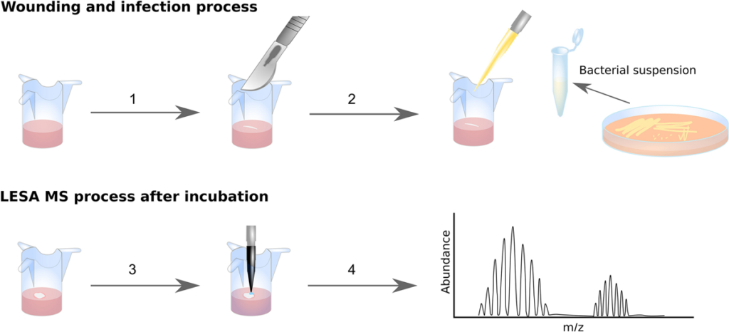
Trauma is one of the leading causes of death in people under the age of 49 and complications due to wound infection are the primary cause of death in the first few days after injury. The ESKAPE pathogens are a group of bacteria that are a leading cause of hospital-acquired infections and a major concern in terms of antibiotic resistance. Here, we demonstrate a novel and highly accurate approach for the rapid identification of ESKAPE pathogens (Enterococcus faecium, Staphylococcus aureus, Klebsiella pneumoniae, Acinetobacter baumannii, Pseudomonas aeruginosa, and Enterobacter spp.) directly from infected wounds in 3D in vitro skin models. Wounded skin models were inoculated with bacteria and left to incubate. Bacterial proteins were identified within minutes, directly from the wound, by liquid extraction surface analysis mass spectrometry. This approach was able to distinguish closely related strains and, unlike genomic approaches, can be modified to provide dynamic information about pathogen behaviour at the wound site. In addition, since human skin proteins were also identified, this method offers the opportunity to analyse both host and pathogen biomarkers during wound infection in near real-time.
University of Cambridge, Royal Holloway University of London
Highlights
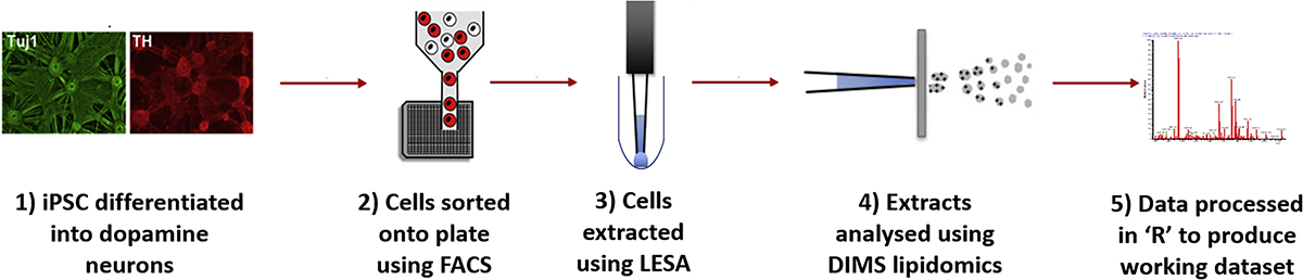
Combining FACS and LESA-MS to establish high-throughput single cell lipid profiling
Lipid differences found within and between populations of human dopamine neurons
Inter-cell lipid heterogeneity is increased in SNCA-A53T dopamine neurons
Identification and isolation of human iPSC-dopamine neurons with a TH-RFP reporter
Summary
Advances in single cell genomics and transcriptomics have shown that at tissue level there is complex cellular heterogeneity. To understand the effect of this inter-cell heterogeneity on metabolism, it is essential to develop a single cell lipid profiling approach that allows the measurement of lipids in large numbers of single cells from a population. This will provide a functional readout of cell activity and membrane structure. Using liquid extraction surface analysis coupled with high-resolution mass spectrometry we have developed a high-throughput method for untargeted single cell lipid profiling. This technological advance highlighted the importance of cellular heterogeneity in the functional metabolism of individual human dopamine neurons, suggesting that A53T alpha-synuclein (SNCA) mutant neurons have impaired membrane function. These results demonstrate that this single cell lipid profiling platform can provide robust data that will expand the frontiers in biomedical research.
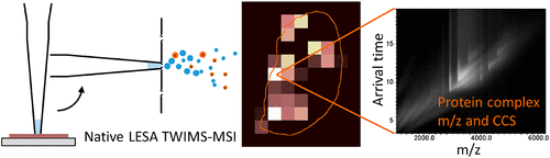
We have previously demonstrated native liquid extraction surface analysis (LESA) mass spectrometry imaging of small intact proteins in thin tissue sections. We also showed calculation of collision cross sections for specific proteins extracted from discrete locations in tissue by LESA traveling wave ion mobility spectrometry (TWIMS). Here, we demonstrate an integrated native LESA TWIMS mass spectrometry imaging (MSI) workflow, in which ion mobility separation is central to the imaging experiment and which provides spatial, conformational, and mass information on endogenous proteins in a single experiment. The approach was applied to MSI of a thin tissue section of mouse kidney. The results show that the benefits of integration of TWIMS include improved specificity of the ion images and the capacity to calculate collision cross sections for any protein or protein complex detected in any pixel (without a priori knowledge of the presence of the protein).
University of North Texas, Huazhong Agricultural University, Heinrich Heine University, University of Goettingen, Leibniz Institute of Plant Genetics & Crop Plant Research, USDA-ARS
Abstract
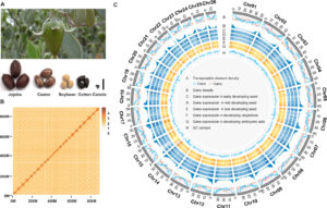
Seeds of the desert shrub, jojoba (Simmondsia chinensis), are an abundant, renewable source of liquid wax esters, which are valued additives in cosmetic products and industrial lubricants. Jojoba is relegated to its own taxonomic family, and there is little genetic information available to elucidate its phylogeny. Here, we report the high-quality, 887-Mb genome of jojoba assembled into 26 chromosomes with 23,490 protein-coding genes. The jojoba genome has only the whole-genome triplication (γ) shared among eudicots and no recent duplications. These genomic resources coupled with extensive transcriptome, proteome, and lipidome data helped to define heterogeneous pathways and machinery for lipid synthesis and storage, provided missing evolutionary history information for this taxonomically segregated dioecious plant species, and will support efforts to improve the agronomic properties of jojoba.
NanoESI-MS/MS and UPLC-nanoESI-MS/MS methods using the Advion TriVersa NanoMate were used to analyze triacylglycerols from jojoba.
Authors: Cecilie Rosting, Jinglei Yu, and Helen J. Cooper*
Abstract
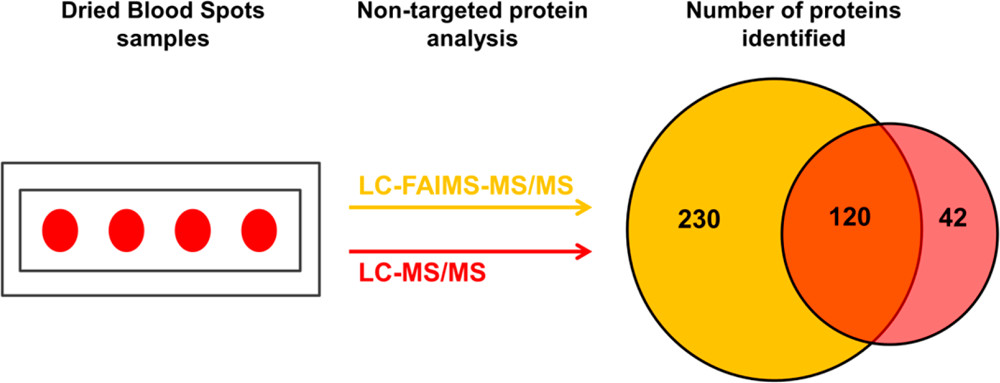
Despite the great potential of dried blood spots (DBS) as a source of endogenous proteins for biomarker discovery, the literature relating to nontargeted bottom-up proteomics of DBS is sparse, primarily due to the inherent complexity and very high dynamic range associated with these samples. Here, we present proof-of-concept results in which we have coupled high field asymmetric waveform ion mobility spectrometry (FAIMS) with liquid chromatography–tandem mass spectrometry (LC–MS/MS) for nontargeted bottom-up proteomics of DBS with the aim of addressing these challenges.
We, and others, have previously demonstrated the benefits of FAIMS more generally in proteomics including improved signal-to-noise and extended proteome coverage, and the aim of the current work was to extend those benefits specifically to DBS. The DBS samples were either extracted by the more traditional manual “punch and elute” approach or by an automated liquid surface extraction (LESA) approach prior to trypsin digestion. The resulting samples were analyzed by LC–MS/MS and LC–FAIMS–MS/MS analysis. The results show that the total number of proteins identified increased by ∼50% for the punch and elute samples and ∼45% for the LESA samples in the LC–FAIMS–MS/MS analysis. For both the punch and elute samples and the LESA samples, ∼30% of the total proteins identified were observed in both the LC–MS/MS and the LC–FAIMS–MS/MS data sets, illustrating the complementarity of the approaches.
Overall, this work demonstrates the benefits of inclusion of FAIMS for nontargeted proteomics of DBS.
Liquid extraction surface analysis (LESA) is an ambient surface sampling technique that allows the analysis of intact proteins directly from tissue samples via mass spectrometry. Integration of ion mobility separation to LESA mass spectrometry workflows has shown significant improvements in the signal-to-noise ratios of the resulting protein mass spectra and hence the number of proteins detected. Here, we report the use of a quadrupole–cyclic ion mobility–time-of-flight mass spectrometer (Q-cIM-ToF) for the analysis of proteins from mouse brain and rat kidney tissues sampled via LESA. Among other features, the instrument allows multiple pass cyclic ion mobility separation, with concomitant increase in resolving power. Single-pass experiments enabled the detection of 30 proteins from mouse brain tissue, rising to 44 when quadrupole isolation was employed. In the absence of ion mobility separation, 21 proteins were detected in rat kidney tissue including the abundant α- and β-globin chains from hemoglobin. Single-pass cyclic ion mobility mass spectrometry enabled the detection of 60 additional proteins. Multipass experiments of a narrow m/z range (m/z 870–920) resulted in the detection of 24 proteins (one pass), 37 proteins (two passes) and 54 proteins (three passes), thus demonstrating the benefits of improved mobility resolving power.
Helen J. Cooper, Emma K. Sisley, Jakub Ujma, Martin Palmer, Kevin Giles, Francisco A. Fernandez-Lima
Shanghai University of Traditional Chinese Medicine
Objective
To establish a simple, rapid and non‐destructive technique for identifying the authenticity of agarwood.
Methods
Liquid extraction surface analysis mass spectrometry (LESA‐MS) was firstly proposed to identify the authenticity of 62 agarwood samples without sample preparation. In addition, multivariate statistical models and thin‐layer chromatography (TLC) method were used to analyse and verify the results of LESA‐MS.
Conclusion
The proposed LESA‐MS method was successfully applied in the direct qualitative analysis of agarwood from different sources. This study indicated great feasibility and practicality of LESA‐MS in the rapid identification of agarwood, and provided a non‐destructive and meaningful preliminary screening tool for the agarwood industry.
The brain is a remarkably complex organ and cholesterol homeostasis underpins brain function. It is known that cholesterol is not evenly distributed across different brain regions; however, the precise map of cholesterol metabolism in the brain remains unclear. If cholesterol metabolism is to be correlated with brain function it is essential to generate such a map. Here we describe an advanced mass spectrometry platform to reveal spatial cholesterol metabolism in situ at 400-µm spot diameter on 10-µm tissue slices from mouse brain. We mapped, not only cholesterol, but also other biologically active sterols arising from cholesterol turnover in both wild type and mice lacking cholesterol 24S-hydroxylase (CYP46A1), the major cholesterol metabolizing enzyme.
Shanghai University of Traditional Chinese Medicine
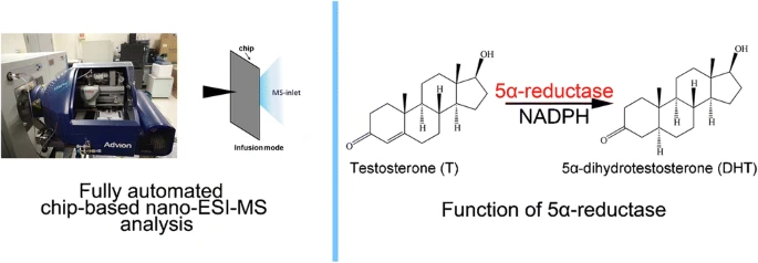
Abstract
The 5α-reductase converts testosterone to dihydrotestosterone (DHT), and excess DHT could cause androgen-related diseases such as androgenetic alopecia and benign prostatic hyperplasia (BPH). To discover new 5α-reductase inhibitors, effective drug screening method with high throughput is thus essential. In this study, fully automated chip-based nanoelectrospray ionization-mass spectrometry (nano-ESI-MS) was innovatively utilized as a screening tool for 5α-reductase inhibitory assay in direct infusion mode, which simplified sample pretreatment and greatly improved experimental efficiency. The preliminary data indicated that curcumin, a natural anti-inflammatory compound, exhibited notably 5α-reductase inhibition activity. Moreover, the obtained results of the chip-based nano-ESI-MS were well consistent with those of HPLC-MS, which suggested that the chip-based nano-ESI-MS could be treated as a rapid and high-throughput drugs screening strategy in pharmaceutical development.